
With regards to diagnosing issues with a canine’s pelvic district, radiographs can be an important instrument. Canine pelvis radiographs can give veterinarians itemized pictures of the bones and joints nearby, assisting them with distinguishing any irregularities or pathologies. In this blog entry, we will investigate the nuts and bolts of canine pelvis radiography, the parts of the canine pelvis that should be visible in radiographs, normal pathologies that can recognized, tips for deciphering these pictures, and progressions in radiographic imaging of the canine pelvis.
The Nuts and bolts of Canine Pelvis Radiography
Radiography, generally used in veterinary practice, offers a painless strategy for looking at a canine’s inner life systems, explicitly the pelvis. This imaging method utilizes X-beams to catch nitty gritty photos of a canine’s pelvic bones, joints, and the nearby delicate tissues, giving priceless understanding to analyze. For the canine pelvis, the cycle includes situating the canine in a manner that enhances the perceivability of the pelvic district on the radiograph.
- Appropriate sedation or sedation is many times important to guarantee the creature stays as yet during the imaging, limiting the possibilities of obscured pictures. The subsequent radiographs manage the cost of veterinarians a reasonable perspective on the pelvic design, including any likely cracks, bone peculiarities, or joint issues. These pictures are essential for distinguishing conditions that could impede a canine’s versatility or cause uneasiness. Perceiving the meaning of these radiographs, veterinarians depend on them to figure out a compelling treatment plan custom-made to the particular requirements of their canine patients. The exact translation of these pictures, contrasting them and laid out standards of canine pelvic life systems, is central for the effective recognizable proof of different pathologies.
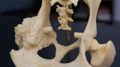
Parts of the Canine Pelvis in Radiographs
In the itemized imaging given by canine pelvis radiographs, veterinarians can intently analyze a few critical physical components. The hip bones, epitomizing the ilium, ischium, and pubis, make the pelvic design supporting the canine’s rump. These components are vital in surveying the general respectability and arrangement of the pelvis. Furthermore, the sacrum and coccygeal vertebrae, which are vital pieces of the spine’s base, show up unmistakably in these pictures. Their condition is crucial for assessing spinal wellbeing and its association with the pelvis.
- The femurs, or thigh bones, act as the interfacing underlying parts between the hip bones and the knees, assuming an essential part in portability. By examining these components in radiographs, veterinarians gain experiences into the canine’s pelvic wellbeing, searching for indications of primary oddities or likely wounds. It is through grasping the typical show of these parts that peculiarities, like misalignments or strange bone thickness, can recognized. This itemized assessment is fundamental for guaranteeing the prosperity of the canine’s portability and by and large wellbeing, working with the early identification and treatment of expected issues.
The Hip Joint: A More critical Look
Vital to a canine’s capacity to move and work, the hip joint is a basic concentration in canine pelvis radiography. This ball-and-attachment joint works with a scope of developments fundamental for running, hopping, and everyday exercises. Radiographic investigation of the hip joint includes a point by point assessment of the femoral head’s fit inside the hip bone socket — the attachment part of the joint. Such examination is vital to recognizing hip dysplasia, a condition set apart by a free fit that can prompt joint unsteadiness, torment, and in the end joint pain.
- Other than dysplasia, radiographs of the hip joint additionally consider the recognition of osteoarthritis, obvious through changes in joint space and the presence of osteophytes or bone spikes. Also, the presence of any delicate tissue expanding or calcification around the joint can allude to incendiary circumstances or injury. Cautious correlation of the hip joint’s radiographic appearance to the laid out standards empowers veterinarians to pinpoint deviations demonstrative of fundamental issues. This scientific cycle not just guides in the early acknowledgment of hip-related pathologies yet additionally directs the improvement of an extensive administration plan custom fitted to lighten the canine’s condition and work on its personal satisfaction.
Normal Pathologies Recognized in Canine Pelvis Radiographs
Radiographs of the canine pelvis are instrumental in uncovering a scope of pathologies that might think twice about canine’s wellbeing and versatility. Among the as often as possible analyzed conditions, breaks present an unmistakable test, noticeable as disturbances or discontinuities in the bone respectability. Disengagements, as well, are promptly obvious on radiographs, where the arrangement of bones, especially at joints, veers off from the ordinary physical situating. Joint inflammation, a typical disease in canines, appears through changes in joint spaces and the presence of osteophytes, flagging joint degeneration and uneasiness.
- Growths inside the pelvic locale, albeit differed by all accounts, can distinguished as strange masses or areas of bone annihilation, requiring further examination to decide their inclination and degree. Formative irregularities, like those found in hip dysplasia, portrayed by a jumble in the fitting of the femoral head to the hip bone socket, demonstrating a potential for future joint issues and agony. Every pathology presents remarkable radiographic highlights, directing the clinician towards an exact conclusion and educating the ensuing course regarding treatment. Understanding the run of the mill show of these circumstances in radiographs is fundamental for veterinarians to really distinguish and resolve the hidden issues influencing the canine pelvis, consequently upgrading the nature of care gave to their four-legged patients.

Deciphering Canine Pelvis Radiographs: Tips and Deceives
Dominating the translation of canine pelvis radiographs relies on the veterinarian’s capacity to recognize the subtleties of ordinary versus obsessive discoveries. A precise methodology is fundamental for a complete assessment. Beginning with bone arrangement, investigate the pelvic symphysis, sacroiliac joints, and the connection between the femoral heads and hip bone socket. Anomalies here could demonstrate disengagement, cracks, or formative issues like hip dysplasia. Moving to bone thickness, varieties might recommend fundamental circumstances like osteoarthritis or neoplastic cycles. It’s essential to analyze the encompassing delicate tissues for indications of enlarging or calcification that could allude to injury or infection.
- A skilled translation likewise includes a next to each other correlation with standard physical references to detect deviations. Information on normal neurotic introductions improves the clinician’s capacity to distinguish inconspicuous changes that might be early signs of sickness. Rehearsing on various cases can construct certainty and capability in recognizing harmless varieties and clinically critical discoveries.
- Veterinarians ought to stay careful for curios – irregularities presented during the imaging system, which could confused with pathologies. Perceiving these requires experience with the gear and imaging strategy utilized. At last, a point by point evaluation joined with an all encompassing perspective on the patient’s clinical picture directs the symptomatic cycle, guaranteeing precise understanding of canine pelvis radiographs.
Progressions in Radiographic Imaging of the Canine Pelvis
The field of veterinary analytic imaging has seen critical mechanical advancement, straightforwardly improving our capacity to investigate the canine pelvis with more noteworthy accuracy and detail. Computerized radiography stands apart by offering speedy handling times and better picture quality analyzed than customary film radiography.
- This jump forward smoothes out the symptomatic cycle as well as decreases the openness to radiation for the two patients and veterinary experts. Further enhancing our demonstrative weapons store, registered tomography (CT) checks present a game-changing benefit by giving three-layered pictures of the pelvic region. This takes into consideration an unrivaled perspective on the intricacy of pelvic wounds or irregularities, working with a thorough comprehension that was beforehand hard to accomplish with two-layered radiographs alone.
- Additionally, attractive reverberation imaging (X-ray) offers an excellent perspective on delicate tissue structures encompassing the pelvic bones. This is especially advantageous for diagnosing conditions that basically influence the delicate tissues, like tendon wounds, without depending exclusively on the more intrusive careful investigations. The capacity of X-ray to separate between different sorts of tissues makes it priceless for point by point assessments of the canine pelvis.
- These progressions have not just pushed the field of veterinary medication into another time of demonstrative ability yet in addition highlight the significance of proceeding with schooling for veterinary experts. Staying informed concerning these mechanical upgrades guarantees that they can use the full range of analytic apparatuses accessible, prompting more exact analyses and custom-made medicines for canine pelvic circumstances.
Conclusion
In synopsis, the utilization of radiographs for looking at the canine pelvis has demonstrated vital for veterinary experts. This symptomatic device considers a nitty gritty perspective on the pelvis, helping with the recognizable proof and comprehension of different circumstances that might influence a canine’s wellbeing and versatility. Through cautious investigation of these pictures, veterinarians can distinguish irregularities, analyze conditions, and foster powerful treatment plans.


One Comment