Understanding the pelvis life structures of canines is vital for diagnosing and treating different muscular circumstances. Radiographic imaging assumes an imperative part in envisioning the designs of the canine pelvis and recognizing any irregularities. In this blog entry, we will investigate the essentials of canine pelvis life systems, the most common way of getting and perusing pelvis radiographs, normal circumstances analyzed through this imaging method, and the headways in radiology for canine muscular consideration.
Prologue to Radiographic Imaging of the Canine Pelvis
Radiographic imaging, frequently alluded to as X-beam innovation, is a basic symptomatic strategy in veterinary medication for investigating the inner life systems of creatures, including canines. This imaging methodology is especially fundamental for inspecting the canine pelvis, offering veterinarians an unmistakable, inside take a gander at the skeletal system and delicate tissue designs of this perplexing region.
- The interaction includes the utilization of X-beams to make point by point pictures of the pelvis, featuring both bone and delicate tissue contrast. These pictures are imperative for surveying the state of the pelvic bones, including the hip joints, sacrum, and encompassing tissues. The capacity to imagine these inner designs with accuracy is basic for recognizing wounds, illnesses, or any formative issues that may not be clear through outer assessment alone.
- Through the essential utilization of radiographic imaging, veterinarians can pinpoint explicit anomalies inside the pelvic district, working with exact conclusions and illuminating resulting treatment methodologies. This procedure requires ability both in the procurement of the pictures, to guarantee they offer the absolute most enlightening perspective, and in their translation, as understanding the subtleties of what is ordinary versus strange is fundamental for viable veterinary consideration.
The Essential Life structures of the Canine Pelvis
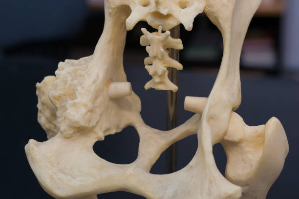
The canine pelvis is a complex and unpredictably organized district that serves numerous capabilities, including supporting the heaviness of the body and working with development. At its center, the pelvis comprised of a few bones that interface with structure a stable, yet adaptable, system. These bones incorporate the ilium, ischium, and pubis, which combine to make the hip bone socket – the attachment that obliges the femoral head, framing the hip joint. This joint is a basic component in the canine’s versatility, considering an extensive variety of movement while likewise bearing the creature’s weight during exercises.
- Notwithstanding these essential bones, the pelvis consolidates the sacrum and coccygeal vertebrae, which anchor the spinal segment to the pelvic area. This association is fundamental for the transmission of powers between the rear appendages and the spine during development and gives a solid groundwork to strong connection and the security of inward organs.
- The design is additionally built up by an organization of tendons and muscles, which support the pelvic bones as well as add to the canine’s train capacities. The encompassing veins and nerves supply the important supplements and signals for the wellbeing and capability of the pelvic district. Understanding this mind boggling life systems is crucial for precise translation of radiographs and the successful finding and treatment of pelvic circumstances in canines.
Setting up Your Canine for a Pelvis Radiograph
At the point when it comes time for your canine ally to go through a pelvis radiograph, guaranteeing their solace and wellbeing is principal. Readiness begins at home, where fasting might prescribed for a few hours preceding the arrangement to lessen the gamble of sedation related confusions. At the veterinary facility, an intensive assessment of your canine’s wellbeing history and an actual assessment will go before any imaging. Sedation or general sedation is usually utilized to keep your canine fixed and torment free during the strategy. This not just works with the procurement of great pictures yet additionally limits pressure for your pet.
- The veterinary group will cautiously situate your canine on the radiography table, really focusing on the arrangement of the rear appendages and pelvis to ensure that the subsequent pictures are pretty much as enlightening as could expected. It’s fundamental for proprietors to comprehend that while the planning and strategy could appear to be overwhelming, these means taken with absolute attention to detail to guarantee the security and prosperity of your pet. A definitive objective is to catch clear, demonstrative quality pictures that will support the exact finding and compelling treatment of any hidden circumstances influencing the pelvis.
Perusing a Canine Pelvis Radiograph: Why Vets Look
While dissecting a canine pelvis radiograph, veterinary experts fastidiously look at the subtleties to distinguish any signs that could show injury or infection. The emphasis is principally on the primary respectability and arrangement of the pelvic bones, which incorporate the ilium, ischium, pubis, and the sacrum, as well as the situating and state of the hip joints. A sharp eye kept on the thickness of the bone designs; varieties here can recommend issues like osteoporosis or malignant growth. Joint spaces assessed for consistency and evenness, where irregularities might flag conditions like joint inflammation or hip dysplasia. The presence of bone prods or uncommon developments is additionally investigated, as these can be demonstrative of degenerative sicknesses or past wounds. Also, the radiographs are painstakingly evaluated for any proof of cracks or separations, which are basic for prompt and suitable administration.
- Past the actual bones, veterinarians additionally look at the encompassing delicate tissue structures for indications of enlarging, masses, or different inconsistencies that could highlight hidden pathology. Regard for these subtleties permits veterinarians to gather an exhaustive comprehension of the canine’s pelvis wellbeing, directing them towards a precise determination and a viable treatment plan. This point by point approach guarantees that nothing ignored, empowering the conveyance of designated care to address the particular requirements of every canine patient.
Normal Circumstances Analyzed Through Pelvis Radiographs
Pelvis radiographs act as a basic demonstrative device in distinguishing different muscular issues in canines. Among the most often analyzed conditions is hip dysplasia, quite predominant in bigger variety canines, where early recognition through radiography can essentially affect the executives and results. Pelvic cracks and femoral breaks are likewise detectable through these nitty gritty pictures, taking into consideration immediate and exact intercession.

- Separations of the hip joint, frequently coming about because of injury, are plainly noticeable on radiographs, empowering veterinarians to quickly devise a strategy for realignment or medical procedure if vital. Also, degenerative joint sickness, including osteoarthritis, can determined to have the guide of pelvis radiographs. This condition’s progressive mileage on joint designs are carefully itemized in radiographic imaging, working with early administration systems to mitigate torment and further develop versatility. Besides, radiographs can uncover bone growths or indications of metastatic illness, directing further demonstrative and remedial activities. From the perspective of radiographic imaging, these normal yet significant circumstances uncovered, considering focused on and viable treatment approaches for canine patients.
The Job of Radiology in Canine Muscular Medical procedure
Radiology is instrumental in the preparation and execution of muscular medical procedures on canines, offering a window into the complicated designs of the canine pelvis before a specialist makes the principal cut. Through itemized pre-usable radiographs, careful groups can acquire significant experiences into the points of interest of the injury or infection influencing the canine’s pelvis or hip joints. This preliminary step is fundamental for planning a careful technique as well as for anticipating and getting ready for potential difficulties that could emerge during the methodology.
- During the medical procedure itself, fluoroscopy, a high level radiographic method that gives ongoing X-beam pictures, turns into a fundamental apparatus. It directs the specialist’s hands, guaranteeing the exact position of screws, plates, or prosthetic parts, which is pivotal for the outcome of the medical procedure and the canine’s recuperation.
- After the medical procedure, the job of radiology reaches out into the post-usable stage, where radiographs are again utilized to survey the result of the methodology. These pictures affirm whether the expected adjustment or fix has accomplished and consider the checking of the recuperating system. They act as a basic designated spot to guarantee that the way to recuperation is on course and that any changes in post-employable consideration made quickly to help the canine’s re-visitation of wellbeing. Through every one of these stages, radiology stays a foundation of canine muscular medical procedure, empowering accuracy, guaranteeing wellbeing, and working with effective results.
Propels in Radiographic Imaging for Canine Pelvis
The domain of radiographic imaging has seen extraordinary development, especially helping the determination and treatment of canine pelvic circumstances. With the coming of advanced radiography, veterinary experts presently appreciate admittance to pictures of predominant clearness and detail. This innovation improves analytic precision as well as fundamentally reduces the time expected to get and deal with these urgent pictures. The advancement doesn’t stop here; figured tomography (CT) filters and attractive reverberation imaging (X-ray) have additionally raised the norm of care. These modalities offer unmatched perspectives on the pelvis in three aspects, uncovering subtleties of bone and delicate tissue structures that were formerly difficult to recognize. This jump in imaging capacities enables veterinarians to pinpoint the specific idea of pelvic irregularities with more prominent accuracy, clearing the w


One Comment